
Acute Renal Failure Ultrasound
Ultrasound is a non-invasive, immediate tool used to image tissue. It will not penetrate bone (like an X-Ray). So the first step to help you read the ultrasound image is to be familiar with the anatomy that you are imaging. Various body tissues conduct sound differently. Some tissues absorb sound waves while others reflect them.

Introduction to the Lower Extremity Venous Doppler Study Ultrasound, Diagnostic medical
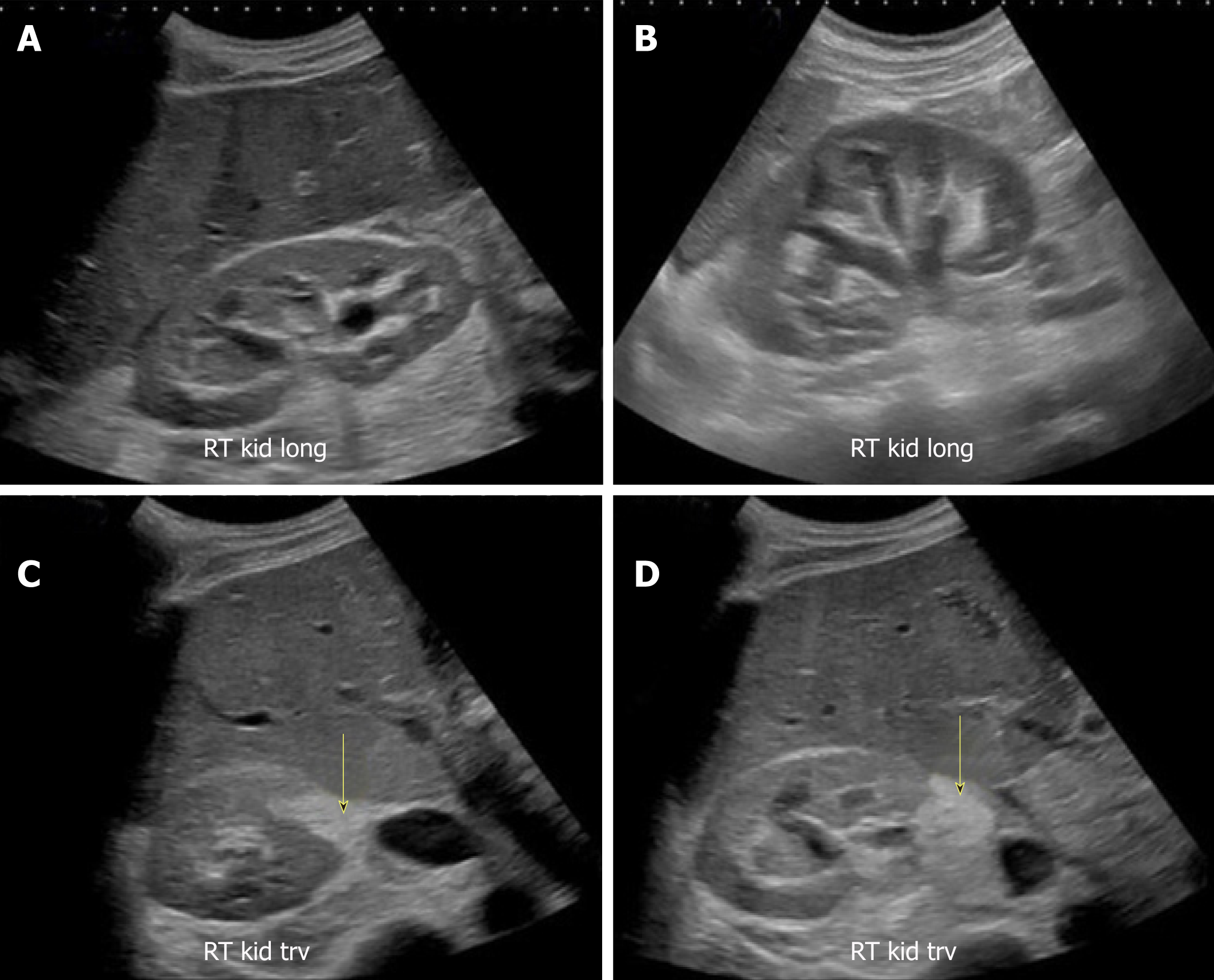
Ultrasonography of the kidneys is essential in the diagnosis and management of kidney-related diseases. The kidneys are easily examined, and most pathological changes in the kidneys are distinguishable with ultrasound. In this pictorial review, the most common findings in renal ultrasound are highlighted. 1.
Cureus OccultPerforated Gangrenous Gallbladder Found on Resonance
For example, say you want to pinpoint the cranial pole of the kidney (red circle): it lies at 10 cm depth.. As we all know, an ultrasound image is made up of lighter and darker areas and several shades of gray. Being the skilled sonographer you are, you already know that this feature of a structure being lighter or darker on the screen is.
Sonoguide // Ultrasound Physics and Technical Facts for the Beginner
Diagnostic ultrasounds use sound waves to make pictures of the body. Ultrasound, also called sonography, shows the structures inside the body. The images can help guide diagnosis and treatment for many diseases and conditions. Most ultrasounds are done using a device outside the body. However, some involve placing a small device inside the body.
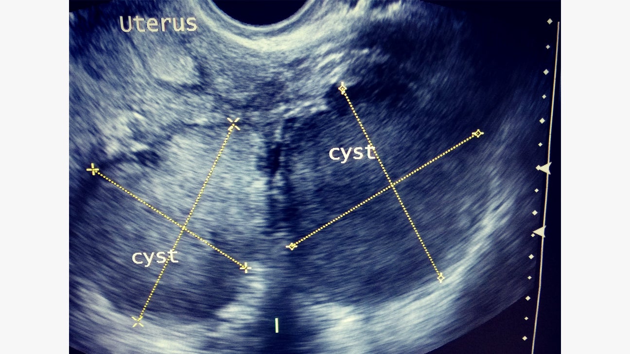
Endometriosis Ultrasound Images
A warm, unscented, hypo-allergenic, water-based ultrasound gel will be applied to the area of concern, and your sonographer will move the transducer around to gather images of your organs. You may be asked to hold your breath and change position to help better examine the area of concern. You may experience mild to moderate pressure while the.
Liver Spots Vs Skin Cancer
The next thing to notice during ultrasound interpretations is the baby. It will look either gray or white on the image. It will be located inside the darker area of the image, which is the amniotic fluid. The amount of detail you can see will depend on the pregnancy state and the development of the baby.

Ultrasound Video showing Acute appendicitis or inflamed appendix. YouTube
Artifacts in Images Artifacts are unwanted elements that appear in ultrasound images, causing distractions and hindering accurate interpretation. They can include speckles, lines, shadows, or other irregular patterns. These artifacts might stem from equipment malfunctions, poor positioning, or incorrect use of ultrasound probes.

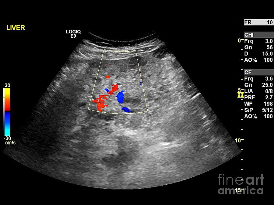
Abnormal results for liver ultrasound what can they mean?
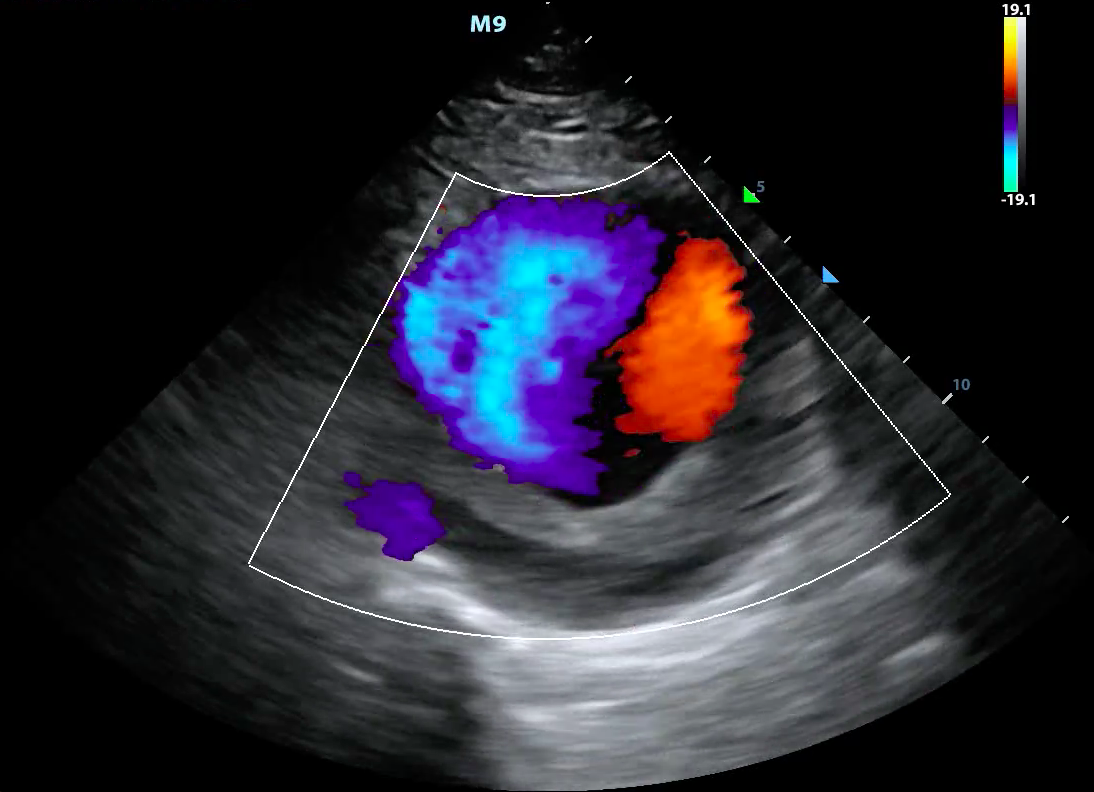
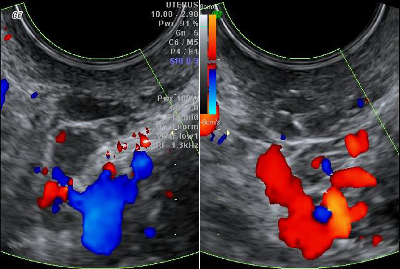
Color-flow imaging (also called triplex ultrasound) is an enhanced form of Doppler ultrasound technology. In a procedure similar to duplex ultrasound, it uses color to highlight the direction of blood flow. Vessels in which blood is flowing are colored red for flow in one direction and blue for flow in the other, with a color scale that.

Abdominal ultrasound reveals a distended gallbladder (9 × 3.8 cm) with... Download Scientific
In conclusion, the meaning of red on ultrasound scans is closely related to blood flow and circulation. Red areas indicate the presence of blood flow and can help identify abnormalities or pathologies. However, accurate interpretation requires proper training, expertise, and consideration of the clinical context.
Microscopic appearance x100 view of left tubal contents showing... Download Scientific Diagram
Essentially, ultrasound uses sound waves to help gather images of different internal parts of your body. These images will be displayed immediately onto a screen and can be saved for a radiologist to interpret later on. Most commonly, ultrasounds are used for pregnancy, diagnosis of soft tissue or organ conditions, medical procedures, and.

Malrotated Kidney Ultrasound YouTube
1. Red. Red color on an echocardiogram represents blood flowing towards the transducer or ultrasound probe. It indicates oxygenated blood flowing from the heart to the rest of the body. Normal red color patterns are essential for healthy blood circulation. 2. Blue. Blue color on an echocardiogram represents blood flowing away from the transducer.
Transvaginal ultrasound JOSEF PFLUG VASCULAR LABORATORY
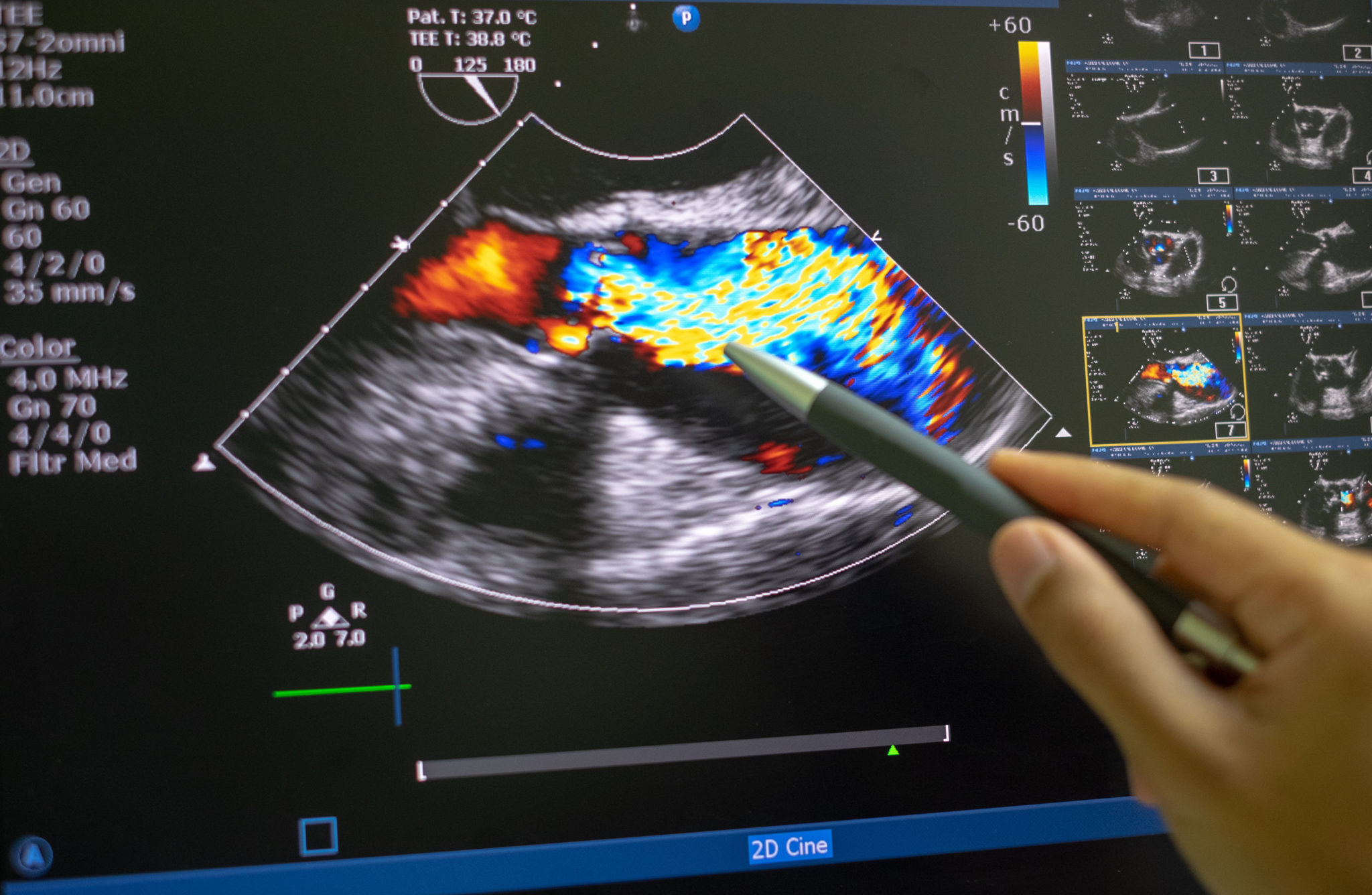
1.8.2.1 Principles of Color Doppler. The advent of color Doppler was a breakthrough in medical ultrasound. With this technique it became possible to directly "observe" blood flow within the heart. Such visualization is achieved by color-encoding Doppler information and displaying the colors as an overlay on the 2D image of the heart.

Ein Ultraschallbild lesen 9 Schritte (mit Bildern) wikiHow
An ultrasound may show gallstones blocking the bile duct. A doctor may also look for gallstones in the gallbladder since this is a common cause of pain in the upper stomach and elevated liver enzymes.

Liver Ultrasound Normal Vs Abnormal Image Appearances Comparison Liver Ultrasound Pathologies
Doppler ultrasound is a specific type of ultrasound exam that is sometimes done with abdominal ultrasound. This helps the radiologist see and evaluate: Blood flow to specific organs. When done with abdominal ultrasound, it is often to look at blood flow to your liver or kidneys. Congenital vascular malformations.

Liver Anatomy and Protocol Medical ultrasound, Liver anatomy, Ultrasound sonography
Atypical liver ultrasound results may include: changes to the size of the liver. extra fat deposits on the liver. lesions or nodules on the liver. scarring on the liver. blockages to the.
Echocardiography (ultrasound). Parker University
Step 2.Understand the Ultrasound Image Orientation. The orientation of the ultrasound is important in order to understand what you see on the screen or printed ultrasound picture. When the radiologist or doctor performs the ultrasound, s/he first checks the probe to identify the indicator.
- Days Until December 31 2023
- Things To See And Do In Oban
- Last Competitor In Relay Team Crossword Clue
- What S The Last Letter Of The Greek Alphabet
- Kaise Mujhe Tum Mil Gayi Serial Written Update
- Enid Graham Movies And Tv Shows
- Calcular Cc De Un Cilindro
- Chinese Restaurants In Newcastle Upon Tyne
- Have A Good Weekend In Spanish
- It Is Well With My Soul Hymn Story